Introduction
The human foot is a marvel of engineering. With 26 bones, over 30 joints, and more than 100 muscles, tendons, and ligaments, it supports our entire body weight while allowing intricate movements. From walking to running, jumping, and even balancing on uneven surfaces, the foot plays a critical role in everyday life. Understanding foot anatomy is essential not only for medical professionals but also for athletes, dancers, and anyone interested in optimizing movement and preventing injuries.
The term foot anatomy encompasses the bones, muscles, ligaments, tendons, and neural networks that allow the foot to function efficiently. From maintaining stability to absorbing shock, the foot is a finely tuned system. This guide provides a comprehensive look at each component, its function, and how it contributes to overall mobility.
Bones of the Foot
The Foot Anatomy comprises 26 bones, categorized into tarsal, metatarsal, and phalangeal bones. These bones form the rigid structure necessary for support and mobility.
Tarsal Bones
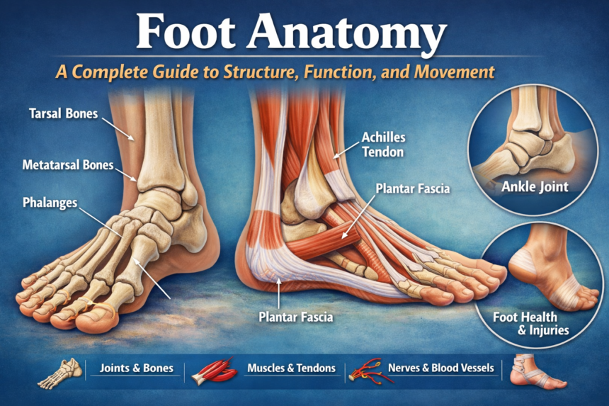
The tarsal bones include the calcaneus (heel), talus (ankle), navicular, cuboid, and three cuneiform bones. The calcaneus bears the majority of weight during standing and walking, while the talus connects the Foot Anatomy to the leg, forming the ankle joint. Together, these bones enable smooth articulation and shock absorption during movement.
Metatarsal Bones
The five metatarsals connect the tarsal bones to the toes, forming the Foot Anatomy midsection. These bones play a crucial role in weight distribution and balance. The first metatarsal, supporting the big toe, carries the highest load during walking and running. Misalignment can lead to conditions like bunions or stress fractures.
Phalanges
The phalanges are the toe bones, with three in each toe except the big toe, which has two. Though small, these bones are essential for balance and propulsion, helping the Foot Anatomy adapt to varied terrain and providing leverage for movements like jumping.
Joints in the Foot
The foot contains several critical joints, enabling a wide range of movements.
Ankle Joint
The ankle joint, or talocrural joint, is a hinge joint connecting the tibia and fibula to the talus. This joint allows dorsiflexion and plantarflexion, essential for walking, running, and climbing stairs. Stability is maintained by surrounding ligaments and tendons, with injuries like sprains being common due to overextension.
Subtalar Joint
Located beneath the ankle, the subtalar joint allows side-to-side motion, facilitating pronation (inward roll) and supination (outward roll). This joint helps absorb shock and adapt to uneven surfaces.
Metatarsophalangeal Joints
The metatarsophalangeal (MTP) joints connect the metatarsals to the phalanges. They provide flexibility for toe movement, essential for push-off during walking and running. Conditions like hammer toe and arthritis often affect these joints.
Muscles of the Foot
Foot muscles are categorized into intrinsic (originating and inserting within the Foot Anatomy ) and extrinsic (originating in the leg).
Intrinsic Muscles
Intrinsic muscles, such as the lumbricals and interossei, are responsible for fine motor control, stabilization, and maintaining the arches of the foot. They ensure balance and aid in precision movements like gripping uneven surfaces.
Extrinsic Muscles
Extrinsic muscles, including the gastrocnemius, soleus, and tibialis anterior, connect the leg to the Foot Anatomy . They generate major forces for walking, running, and jumping. Dysfunction in these muscles can lead to altered gait and increased injury risk.
Ligaments and Tendons
Ligaments and tendons support foot bones and joints, providing stability while permitting mobility.
Plantar Fascia
The plantar fascia runs along the bottom of the Foot Anatomy , forming the plantar arch. It absorbs shock, supports weight, and contributes to the foot’s spring-like function. Overuse can cause plantar fasciitis, a common source of heel pain.
Achilles Tendon
The Achilles tendon connects the calf muscles to the calcaneus. It is critical for pushing off during walking and running. Injuries include tendonitis and ruptures, which can significantly impair mobility.
Arches of the Foot
The Foot Anatomy has three primary arches: medial longitudinal, lateral longitudinal, and transverse. They act as shock absorbers and adapt to uneven terrain. Proper arch support is essential for preventing injuries and maintaining efficient gait mechanics.
Blood Supply and Nerves
The Foot Anatomy vascular system, including the dorsalis pedis and posterior tibial arteries, ensures adequate blood flow. The tibial and peroneal nerves provide sensation and motor control. Circulatory or nerve compromise can lead to severe conditions, including ulcers or neuropathy.
Common Foot Movements
The foot enables walking, running, jumping, and balancing. Movements involve coordinated actions of bones, muscles, and tendons. Efficient motion reduces energy expenditure and prevents overuse injuries.
Foot Health and Injuries
Common issues include bunions, plantar fasciitis, flat feet, stress fractures, and ankle sprains. Preventive care, proper footwear, and targeted exercises maintain foot health.
Foot Anatomy in Different Populations
-
Children: Developing arches and flexibility
-
Adults: Weight-bearing and posture alignment
-
Athletes: Increased stress on joints and tendons requiring specialized care
Foot Anatomy and Footwear
Footwear significantly influences foot anatomy and function. Supportive shoes enhance comfort, prevent deformities, and improve athletic performance. Improper shoes can cause bunions, hammertoes, or plantar fasciitis.
Advanced Imaging and Foot Anatomy
Techniques like X-rays, MRI, and ultrasound allow detailed visualization of bones, joints, and soft tissues. These tools assist in diagnosing injuries and planning interventions.
Rehabilitation and Therapy
Exercises, stretching, and physiotherapy help restore Foot Anatomy function. Common practices include balance training, strengthening exercises, and gait correction, which prevent future injuries and enhance mobility.
FAQs
1. How many bones are in the human foot?
The human Foot Anatomy contains 26 bones categorized into tarsals, metatarsals, and phalanges.
2. What is the function of the plantar fascia?
The plantar fascia supports the arches of the Foot Anatomy , absorbs shock, and aids in walking and running.
3. How does the Achilles tendon affect movement?
It connects calf muscles to the heel, allowing push-off during walking, running, and jumping.
4. What causes flat feet?
Flat feet occur when the medial arch collapses, which can be due to genetics, injury, or overuse.
5. Which muscles stabilize the Foot Anatomy ?
Intrinsic muscles like lumbricals and interossei stabilize the Foot Anatomy , while extrinsic muscles control larger movements.
6. How can I prevent common Foot Anatomy injuries?
Proper footwear, strengthening exercises, stretching, and avoiding overuse reduce injury risk.
Conclusion
The foot is a complex structure vital to mobility and overall health. Understanding foot anatomy—from bones and joints to muscles, ligaments, and nerves—helps prevent injuries, improve performance, and maintain long-term functionality. By combining proper care, awareness, and supportive practices, everyone can ensure their feet stay healthy and strong. For more detailed insights into Foot Anatomy function and therapy, visit American Podiatric Medical Association.